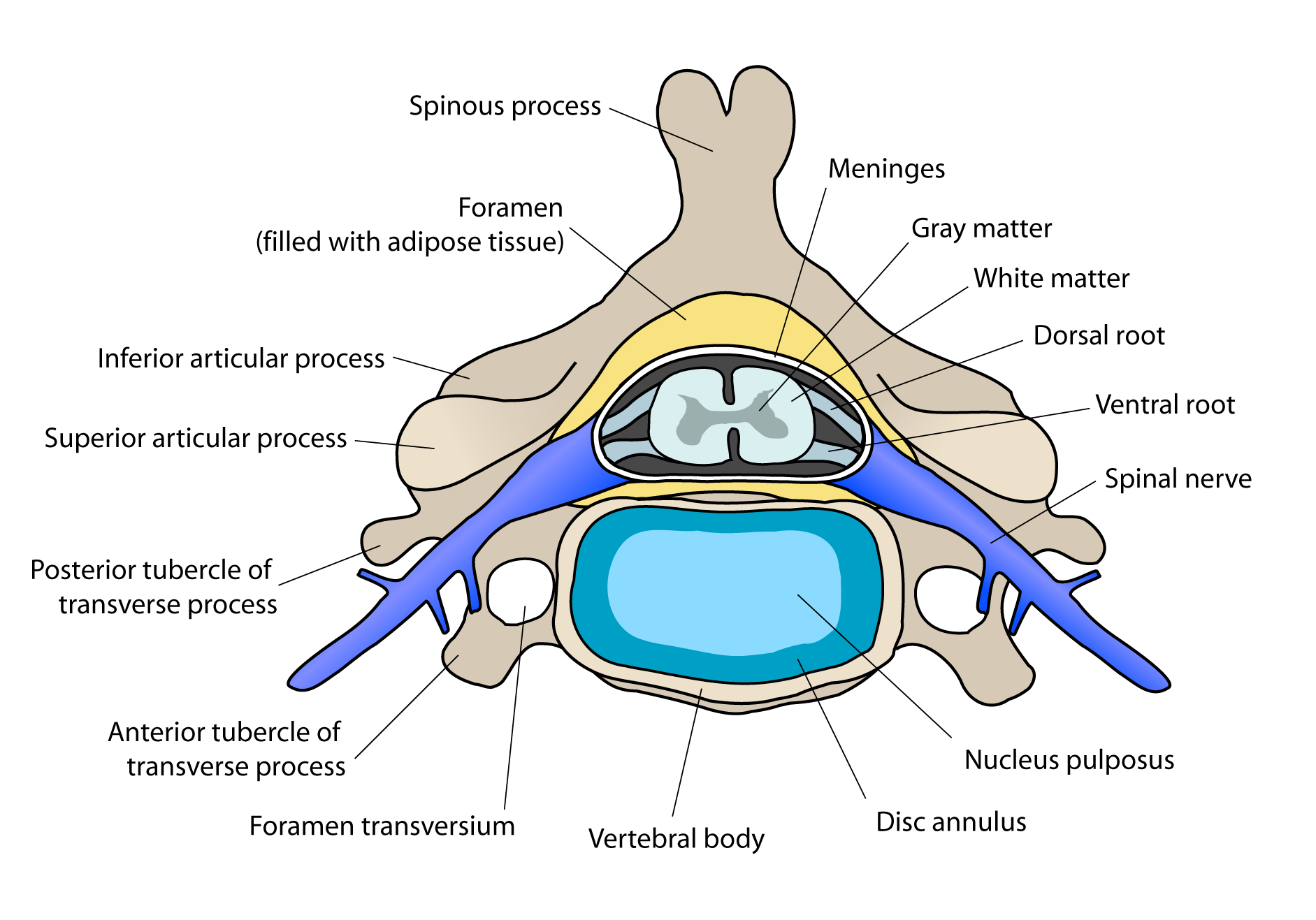
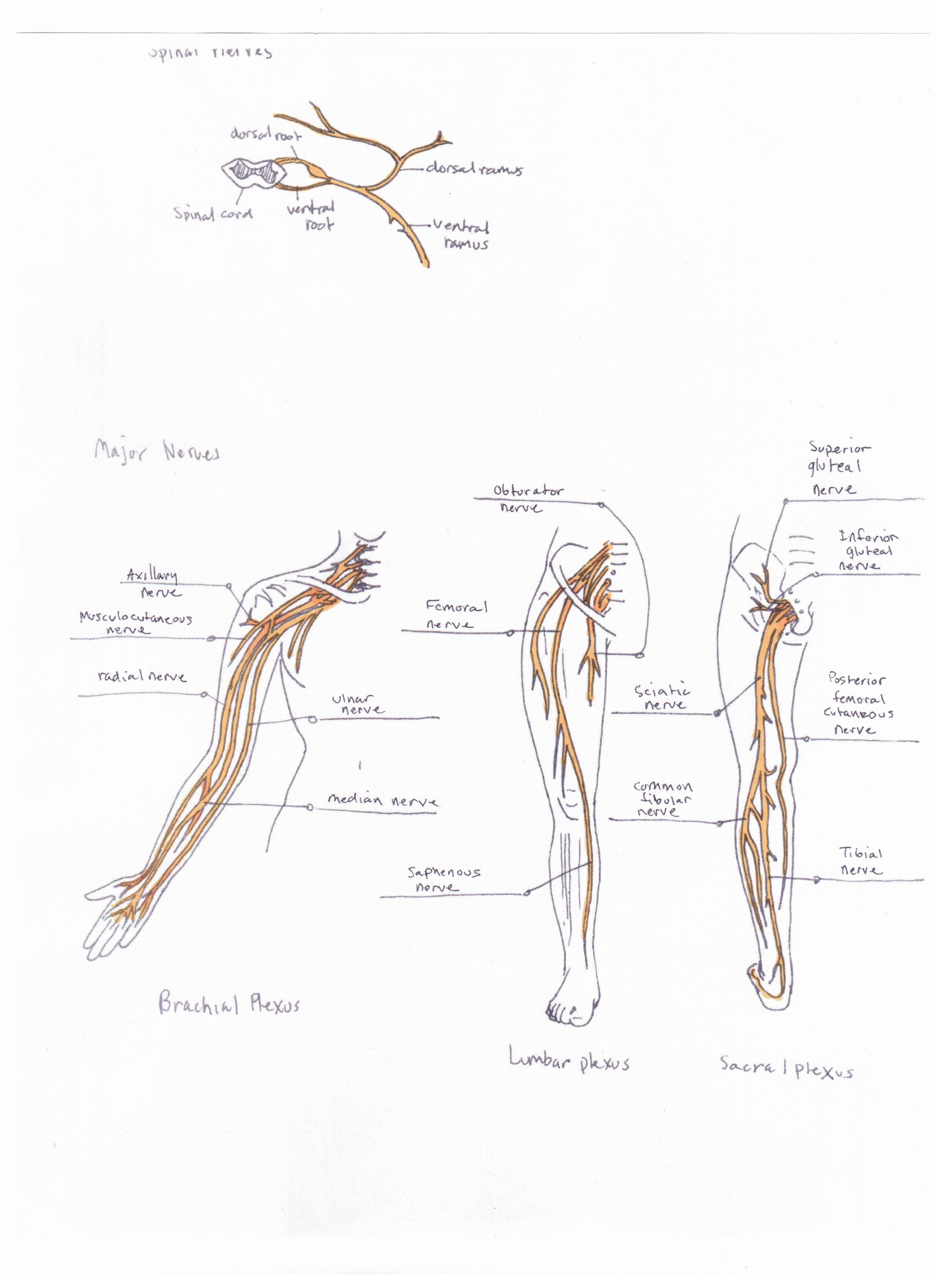
typical spinal nerve diagram
Ventral root - wikidoc. 14 Images about Ventral root - wikidoc : Spinal Nerve - Assignment Point, What Does Ventral Mean In Anatomy and also Typical vertebral hemangioma. Axial (a) and coronal (b) CT images in.
Ventral Root - Wikidoc
 www.wikidoc.org
cervical ventral root vertebra wikidoc
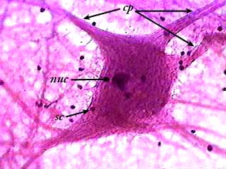
Nervous Tissue
 www.austincc.edu
tissue nervous microscope under neuron motor 400x smear tissues austincc edu
Typical Vertebral Hemangioma. Axial (a) And Coronal (b) CT Images In
 www.researchgate.net
hemangioma vertebral spine mri t2 vertebra incidental coronal syrinx sagittal idiopathic trabeculae bony prominence axiala fig5
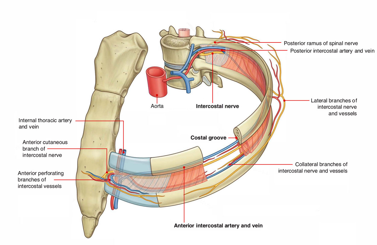
Easy Notes On 【Intercostal Space】Learn In Just 4 Minutes! – Earth's Lab
 www.earthslab.com
intercostal spaces space anatomy anterolateral contents
Print Exercise 17: Histology Of Nervous Tissue Flashcards | Easy Notecards
 www.easynotecards.com
afferent neurons function efferent exercise neuron multipolar nervous histology which nerves sensory interneuron motor identify association unipolar correctly tissue types
Nerve Cell Diagram Labeled 2019 – 101 Diagrams
 www.101diagrams.com
hormonesmatter
Spinal Nerve - Assignment Point
 www.assignmentpoint.com
spinal nerve peripheral nervous system
Spinal Nerve Origin: Cross Sections Exit Of Spinal Nerves Spinal Nerves
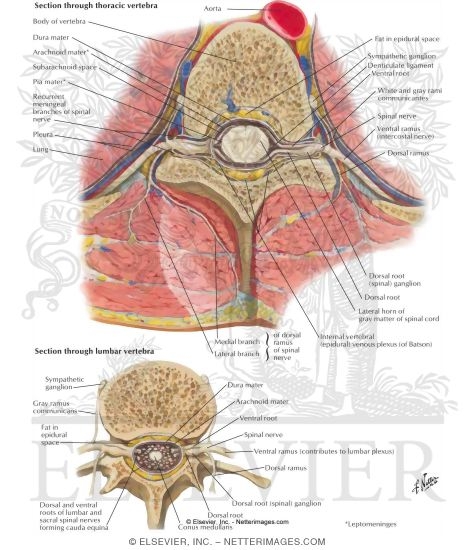 www.netterimages.com
spinal cross nerve origin nerves sections anatomy netter atlas exit slide pricing labeled low netterimages
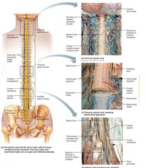
Spinal Nerves | Veterian Key
 veteriankey.com
spinal nerves nerve figure diagram
Chapter 12 - The Spinal Cord Example | Graduateway
 graduateway.com
nerves
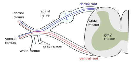
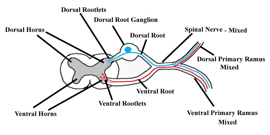
What Does Ventral Mean In Anatomy
 boundbobskryptis.blogspot.com
spinal ventral dorsal ramus nervous efferent
What Is A Spinal Nerve? - Quora
nerve
Anatomy And Physiology - Home
 anatomyscholarsrphs.weebly.com
nerves spinal
Human Physiology - Neurons & The Nervous System II
 people.eku.edu
sympathetic ramus visceral ganglion gray nervous system chain structure cranial drawing neurons mcgill through muscle cardiac ritchisong eku edu
Sympathetic ramus visceral ganglion gray nervous system chain structure cranial drawing neurons mcgill through muscle cardiac ritchisong eku edu. Nerve cell diagram labeled 2019 – 101 diagrams. Spinal nerves nerve figure diagram